
 INTRODUCTION
Excessive load and tension at the point of insertion of plantar fascia lead to inflammatory condition known as plantar fasciitis (PF). Plantar fasciitis is a major cause of the pain at the plantar aspect of the heel; the common site of pain is the medial tubercle of calcaneus (Roxas 2005; David & Lori 2004; Quaschnick 1996; Chandler & Kibler1993). Treatment remedies include both surgical and non-surgical interventions. Non surgical or conservative treatment comprises Non Steroidal Anti Inflammatory Drugs (NSAIDs), stretching, icing, physical therapy and orthotic management including foot orthosis and night splints (AFO), shoe modifications, low dye taping and walking casts (Kavros, 2005; Roxas, 2005; Young, Rutherford & Niedfeldt 2001). The aim of orthotic management is relief pain; reduce stresses on plantar fascia and increase in shock absorption. Also to maintain normal alignment of the foot to allow the fascia to heal. The aim of this paper is to discuss the outcomes of different orthotic devices and techniques for the management of PF in terms of pain relief, resolution of the symptoms and improvement in the anatomical and biomechanical condition.
Signs and Symptoms
Pain is classical symptom of PF, patient feels sever pain when he takes first few steps after sleep or long period of rest. It is sharp, knife like and piercing pain at the medial aspect of the heel and lessens or resolve as patient worm up. Prolong standing will also cause pain along with stiffness. There is localized tenderness of the tissue at the site of the pain. Tenderness can be felt when toes are hyper extended and ankle is dorsiflexed. A calcaneal spur is visible in the X-ray of affected foot (Cole, Craig & Gazewood, 2005; Young, Rutherford & Niedfeldt, 2001).
Etiology And Risk Factor
Etiology of plantar fasciitis is multifactoral, including anatomical, biomechanical and environmental factors. Anatomical factors include flat foot, cavus foot, obesity, and leg length discrepancy and fat pad atrophy.Biomechanical factor are equinus, weak plantar flexors, weak intrinsic muscle and excessive pronation at subtalar joint, whereas trauma, walking bare foot, walking on rough surface for a long time are included in environmental factors (Martin, Hosch, Goforth, Murff, Lynch & Odom, 2001; Gross, Davlin & Evanski 1991).
PATHOLOGY OF PLANTAR FASCIITIS (PF)
Plantar fascia is attached to the medial tubercle of calcaneus proximally and metatarsal head distally. With weight bearing it stretched and micro tears of fascia occur as well as tearing of periosteum at point of attachment of fascia. During rest these micro tears undergo healing and remodeling results in the formation of big bony mass or scar, the heel spur (Young, Rutherford & Niedfeldt, 2001; Quaschnick, 1996; Ryan, 1995; Corrigan & Maitland, 1994). There are inflammatory responses in plantar fascia from disorder of collagen fiber as result of repetition of excessive stress leads to chronic degenerative changes (Stadler, Johnson & Stephens, 2003).
In PF, Plantar flexors strength is decreased and gastro-soleus complex is tightened. Excessive pronation causes instability of hind foot, which leads to more strain on the origin of plantar fascia. Entrapment of medial calcaneal branch of posterior tibial nerve also causes pain. (Roxas 2005; Young, Rutherford & Niedfeldt, 2001; Quaschnick, 1996).
TREATMENT MODALITIES
Both surgical procedures and conservative managements are used to treat plantar fasciitis. Application of ice, use of NSAIDs, stretching and strengthening exercises for gaestrosoleus tightness and plantar flexors strength are non-surgical intervention as well as the orthotic management. Orthotic management is accomplished with use of different types of foot orthoses, heel pads, night splints, low dye taping and walking casts (Kavros, 2005; Sobel, Levitz & Caselli,
1999; Lynch, Goforth, Martin, Odom, Preece & Kotter, 1998).
AIM OF ORTHOTIC MANAGEMENT OF PLANTAR FASCIITIS
PF is self-limiting condition, but it takes a long period usually 6-18 months for complete resolution of symptoms, it is frustrating for both patient and physician. Aim of orthotic management is to address the pain, excessive stresses and inflammatory condition; provide shock absorption and to deal with biomechanical adaptation like slight varus position
of the heel and excessive pronation of fore foot during heel strike that lead to pain in foot and other part of body (Roxas, 2005; Young, Rutherford & Niedfeldt, 2001). Another important goal of the treatment is to keep the plantar fascia in its proper length, during weight bearing and non-weight bearing and restore the strength and flexibility of involved tissues (Barry, Anna & Yinpu. 2002). Injuries to the foot are usually the result of varying degree and size of the kinetic and kinametic changes in the foot, the use of correct orthosis that addresses these changes will result in the relief of the symptoms (Kavros, 2005).

 FOOT ORTHOSES
Foot orthoses are effective in reducing pain in heel and arch area and provide comfort by altering the intrinsic factor and realigning the foot by keeping the sub tarsal joint in neutral position and preventing forefoot pronation, thus improve activity level (Karas &, David, 2002; Sobel, Levitz & Caselli, 1999; Gill & Kiebzak 1996). This goal is also achieved by support the medial longitudinal arch with rigid foundation to reduce the tensile forces within fascia (Gross, Byers, Krafft, Lackey & Melton 2002; Joan & Mark, 2001; Sobel, Levitz & Caselli, 1999; Turlik, Donatalli & Veremis 1999; Lynch et al, 1998).
Prefabricated semi rigid orthoses are effective in pain relief either by realigning the foot or by soft cushioning effect Success rate for foot orthoses is 83-100 % and there is high level of compliance (Kavros, 2005; Landorf, Keenan & Herbert, 2004; Martin et al, 2001; Pfeffer et al, 1999; Gross, Davlin & Evanski, 1991).

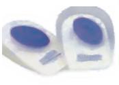 Viscoelastic Heel Pads
Viscoelastic heel pads and cushions are used to replace the atrophied fat pads of the heel and act as shock absorber during heel strike to dissipate the ground reaction forces hence reducing the impact force on leg and spine. Silicon heel pads provide relief at the site of tenderness and heel spur by distributing the weight around the point. Rear foot varus post or lateral wedge reduces pronation of forefoot and reduce the stretch to fascia result in the resolution of symptoms specially the pain (Landorf, Keenan & Herbert, 2004; Seligman & Dawson, 2003; Caselli, 1999; Pfeffer et al 1999).
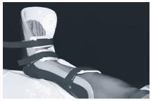 Night Splints
During sleeping irrespective of whether supine or prone position, foot is postured in plantar flexion due to normal tone of gastrosoleus complex, this non functional plantar flexion will result in tightness of Achilles tendon and plantar fascia. Patient will experience a sharp pain at heel when foot touches the ground during first step in the morning due to stretching of tight plantar fascia. Tension night splint in chronic PF keeps foot in dorsi flexion preventing the contraction of the plantar fascia and Achilles tendon, which relaxed in stretched position due to stress relaxation, thus proper length of fascia is maintained (Barry, Anna & Yinpu. 2002; Probe, Baca, Adams & Preece, 1999; Powell, Post, Keener & Wearden, 1998; Chandler & Kibler1993).
The results are supplemented if night splint is used in conjunct with other conservative treatment such as FO, low dye taping and NSAIDs. 100% of success is achieved by use of night splint in chronic cases although these devices have no significant effect during acute phase. Unfortunately few patients feel unpleasant to use AFO during sleep or they feel
discomfort numbness or non compliance (Ryan, 1995; Wapner & Sharkey, 1991).
The night splint made of polypropylene aligned in 5 dorsiflexion and 30 dorsiflexion at metatarsophalangeal joint by placing a wedge under forefoot could be prescribed in chronic cases.
 Low- Dye Taping
Taping is less expensive but more effective way of treatment of the plantar fasciitis, Though it is not considered as proper orthotic technique but still used in initial managements of planter fasciitis. Taping maintain the arch, stabilizes the metatarsal head and pronation of the foot (Cole, Craig & Gazewood, 2005; Quaschnick, 1996; Young, Rutherford & Niedfeldt 2001).
The injuries where there are very little traces of inflammation are present, tissue healing is allowed by protected or very limited ROM through taping during acute phase. Taping is also used as indicator of the Plantar fasciitis if symptoms still persist after few weeks of taping, the patient will reassessed for plantar fasciitis. If it is successful then permanent orthosis is prescribed (Brian, 2006; Landorf, Radford, Keenan, & Remond, 2005; Osbome & Allison, 2006; Chandler & Kibler, 1993).
Supportive Foot Wear
Proper shoes with proper fit and well supported arch and mid sole should be used in case of PF for proper distribution of forces. Shoes must be changed frequently and new shoes can be readjusted according to need, thus improving the symptoms. Shoe with well defined medial arch will reduce the strain in fascia. (Young, Rutherford & Niedfeldt 2001; Quaschnick, 1996; David, 1991).Older shoe will exacerbate the condition in the PF. Plantar fascia will under go tension from heel rise to toe off during gait and will cause the irritation of already inflamed ligament, which can be reduced or even diminished by modifying the commercially available shoes. Such modification include placement medial wedge to elevate arch and placement of silicon pads etc. shoe with proper fit will prevent the recurrence of disorder. (Joan & Mark, 2001; Sobel, Levitz & Caselli, 1999; Mizel, Marymont, & Trepman, 1996).
Walking Casts
In some cases casting is considered to be an effective way of treatment. This process is accomplished by wrapping ankle in neutral position or slightly dorsiflexion with fiberglass walking cast. Walking cast provides rest for heel and reduces the pressure at heel strike. These are also used to provide arch support and avoid the tightness of the Achilles tendon. (Roxas,
2005; Sobel, Levitz & Caselli, 1999; Gill & Kiebzak, 1996).
CONCLUSION
To sum up, PF is one of the common disorders of the foot that causes localized pain in the heel as well as change the functional biomechanics of the patient which in turn affect the activity level of the sufferer. The aim for orthotic management of plantar fasciitis is to deal with pain relief, correction of the posture (excessive pronation and varus heel) and hence the restoring the activities of the patient. Orthotic technique includes the foot orthosis, shoe inserts heel pads,
taping and casting of the foot. No single treatment is thought to be effective. Foot orthosis and heel pads produce relief in the pain by direct cushioning effect or by elevated arch support and controls the pronation of the foot during walking and running thus reduces the tensile forces on the foot, consequently improving the functional restorations. Heel pads also act as shock absorber at heel strike and reduce the overall impact on the leg and low back. In chronic cases the use of night splint aligned in 5 dorsi flexion cause considerable reduction in pain. Night
splints are also effective in the improving the symptoms in recalcitrant PF and reduce the formation of the micro tears. Arch taping and walking casts used to minimize the ROM at ankle during acute phase. Finally, there is no single treatment modality that is effective in the management of the PF; it is combination of the treatments that result in the resolution of the symptoms pain, inflammation and biomechanical factors.
SUGGESTED REFFERENCES:
- Barry, L.D., Anna, N. and Yinpu, C. ( 2002) A retrospective study of standing gastrocnemius-soleus stretching versus night splinting in the treatment of plantar fasciitis. Journal of Foot & Ankle Surgery. 41(4):221-7,
- Brian, F (2006) Plantar Fasciitis: how to maximize outcomes with conservative treatment Podiatry Today, 19 (5), 48 – 56
- Chandler, T . and Kibler, W B. (1993) A biomechanical approach to the prevention, treatment and rehabilitation of plantar fasciitis. Sports Medicine, 15(5):344-52,
- Cole, C.S., Craig, G.J. (2005) plantar fasciitis: evidence-based review of diagnosis and therapy. [Review] American Family Physician, 72(11):2237-42,
- David O. D. (1991) A Comparison of Shoe Inserts to Taping for Painful Arches, Journal of Prosthetics and Orthotics 3(2); 84-. Gill, L H. and Kiebzak, G. M. (1996) Outcome of no surgical treatment for plantar fasciitis Foot & Ankle International. 17(9):527-32,
- Gross, M.T., Byers, J.M., Kraft ,J.L. , Lackey, E.L and Melton K.M. (2002) The impact of custom semi rigid foot orthotics on pain and disability for individuals with plantar fasciitis: Journal of Orthopeadic And sports physical therapy 32(4):149-57
- Joan, M. B. and Mark, W.N. (2001) Over the counter foot remedies Journal of American Family Physician 64(5) 791-6
- Karas, M .A. and David, J.H., (2002) Compensatory Midfoot Dorsiflexion in the Individual with Heel cord Tightness: Implications for Orthotic Device Designs, Journal of Prosthetics and Orthotics 14(2); 82-93.
- http://www.ankle-foot.com accessed on 15 -10-2008
- http://www.icbmedical.com/conditions/heelspurs/low_dye_strapping accessed on 15 -10-2008
KONTAK PEMESANAN KAKI PALSU DAN TANGAN PALSU PT KUSPITO KAKI PALSU:
Website Resmi PT KUSPITO KAKI PALSU : kuspito.com
Call PT KUSPITO KAKI PALSU : 0811263370
Sms PT KUSPITO KAKI PALSU : 0811263370
WhatsApp PT KUSPITO KAKI PALSU : 0811263370
Telegram PT KUSPITO KAKI PALSU : 0811263370
BBM PT KUSPITO KAKI PALSU : KUSPITO
Promo Kaki Palsu Atas Lutut Impor Diskon 50% Cara daftar: SMS KE 085-6000-30000 dengan format: NAMA (SPASI) TFPMODPROMO
Promo Kaki Palsu Bawah Lutut Impor Diskon 50 %
Cara daftar: SMS KE 085-6000-30000 dengan format: NAMA (SPASI) TTPMODPROMO
PT. KUSPITO Ortotik Prostetik adalah Sebuah Perusahaan yang bergerak dibidang ortotik prostetik, memberikan layanan rehabilitasi fisik untuk masyarakat memproduksi alat bantu Prosthesis (kaki palsu, tangan palsu atau kaki tiruan, tangan tiruan) dan Orthosis (alat bantu orthopedi, alat bantu mengkoreksi kecacatan, penyangga kaki yg layuh, dsb).
PT. KUSPITO Ortotik Prostetik tidak hanya bergerak dibidang industri alat bantu ortotik prostetik, melainkan juga bergerak dalam industrik bidang Alat Kesehatan dan Pengadaan Alat kesehatan.
Kantor Pusat PT. KUSPITO Ortotik Prostetik, gedung milik sendiri, gedung warna merah 3 lantai, lokasi pinggir jalan Raya Provinsi yang sangat mudah diakses menggunakan transportasi Umum (Bus) maupun Kendaraan Pribadi. Kuspito Pusat beralamat di Jalan Raya Solo – Tawangmangu KM.12 Papahan Tasikmadu Karanganyar SOLO Jawa Tengah,
bisa dilihat di lokasi Google Map Kuspito Solo
PETA KUSPITO SOLO
Kantor Cabang PT. KUSPITO Ortotik Prostetik di BEKASI, gedung milik sendiri, gedung 1 lantai yang berlokasi di tengah kota, terletak di Pinggir jalan Raya sehingga mudah diakses dengan menggunakan transportasi Umum (Bus) maupun Kendaraan Pribadi. Lokasi Kuspito Bekasi di Jalan Baru Underpass No.4, Duren Jaya, Bekasi Timur, Kota Bekasi , Jawa Barat. (Belakang Rumah Sakit Bella Bekasi)
bisa dilihat di lokasi Google Map Kuspito Bekasi
PETA KUSPITO SOLO
Legalitas PT. Kuspito Ortotik Prostetik
PT . Kuspito Ortotik Prostetik memiliki semua ijin dalam menjalankan usaha. SIUP, TDP, HO, NIK, API, PAK dan semua kebutuhan untuk pengadaan
PT. KUSPITO Ortotik Prostetik bergerak dibidang:
Pembuatan Kaki Palsu / kaki tiruan (Lower Limb Prosthesis)
Pembuatan Tangan Palsu / tangan tiruan (Upper Limb Prosthesis)
Pembuatan Alat Bantu Orthopedi pengkoreksi kecacatan dan disabilitas (Orthosis / Splint / Brace)
Pembuatan Alat bantu Mobilitas
Penjualan Komponen Prosthesis Orthosis
Pengadaan Peralatan maupun produk Alat Kesehatan Ortotik prostetik, Okupasi terapi, Terapi Wicara, Fisioterapi di Instansi dan Rumah Sakit Pemerintah maupun Swasta
Konsultasi masalah Orthopedi
Produk Promo Diskon Unggulan
Promo Kaki Palsu Bawah Lutut Impor Diskon 50 %
Cara daftar: SMS KE 085-6000-30000 dengan format: NAMA (SPASI) TTPMODPROMO
Promo Kaki Palsu Atas Lutut Impor Diskon 50%
Cara daftar: SMS KE 085-6000-30000 dengan format: NAMA (SPASI) TFPMODPROMO
